
Radiology is one of the medical fields where AI has been deployed earliest and most extensively. Three years after the first deployments in public hospitals, it is time to take stock.
What these tools actually do
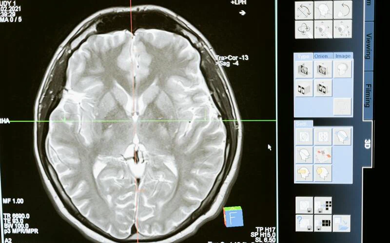
The systems deployed at the university hospitals of Lyon, Bordeaux and Strasbourg focus primarily on two tasks: detecting pulmonary nodules on chest CT scans, and detecting fractures on standard X‑rays.
In both cases, AI plays the role of a safety net. It analyses the image in parallel with the radiologist and raises an alert if it detects something the clinician has not mentioned in their report.
Measured gains
At Bordeaux University Hospital, the detection rate for pulmonary nodules under 6mm increased by 23% since the tool was introduced, according to data published by the thoracic radiology department in 2025.
At Lyon University Hospital, the processing time for radiological emergencies fell by 18% thanks to automatic prioritisation of critical cases in the reading queue.
New constraints
Radiologists interviewed also describe negative effects. The first is the cognitive overload from false alerts. A tool with 95% sensitivity processing 200 examinations per day mechanically generates 10 false alerts daily, each requiring a manual check.
The second constraint is legal: in the event of a dispute, the question of shared liability between the clinician and the system has not yet been settled by French case law.
Key takeaway
AI measurably improves detection, but it also generates additional work. Its net benefit depends heavily on the volume of activity in the department and on the quality of its integration into the clinical workflow.